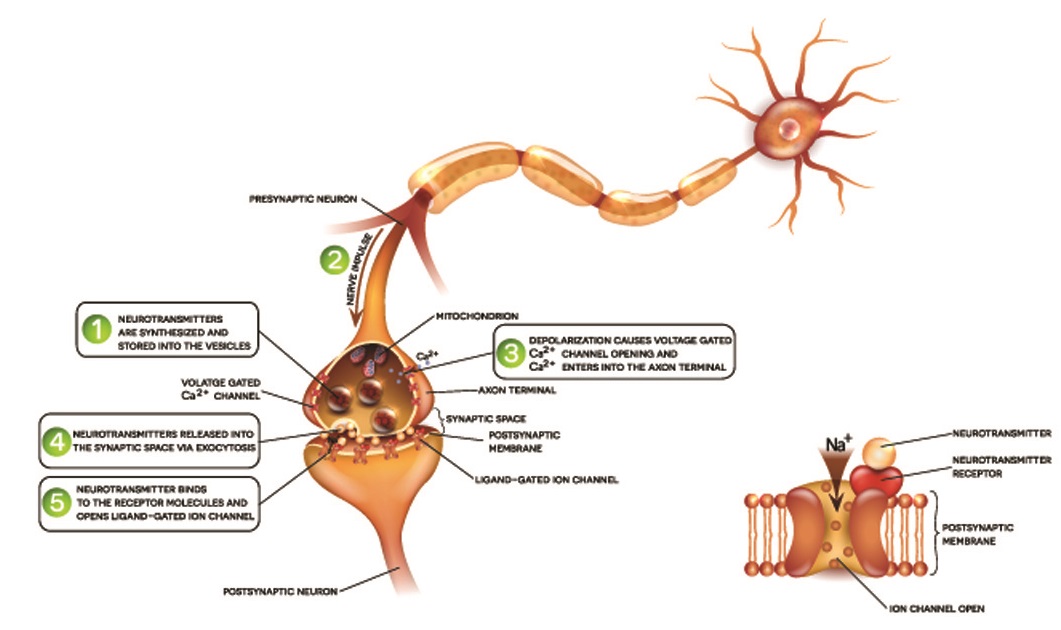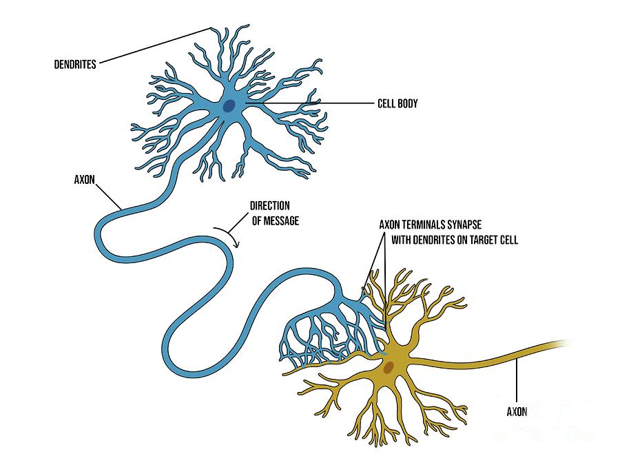


It therefore appears that a novel regenerative pathway underlies Dlk-independent regeneration. Additionally, conventional MAPK genes downstream of Dlk are not required-including the PMK-3/CEBP-1 p38 pathway-nor was the parallel MLK-1/KGB-1/FOS-2 JNK MAPK cascade involved. For example, Dlk-independent regeneration proceeds more slowly compared with conventional regeneration, initiating around 24 h after surgery, compared with ∼12 h in control axons, and this is true in both young and old worms. Importantly, the authors also identify cellular and molecular features that distinguish this form of regeneration from previously described regenerative events. ( 5) show that severing ASJ sensory dendrites in wild-type animals also significantly enhances the axonal regenerative response compared with axotomy-alone controls, arguing against this possibility. Although it is a possibility that such regenerative effects following dendrite lesion are specific to dlk-1 mutants, Chung et al. Unraveling the genetic basis of axonal regeneration has been challenging, so the discovery of a Dlk-independent regenerative event is an important step forward for modeling axonal responses to neuronal injury. How well these axons generate functional connections in the nerve ring remains an open question. The regeneration of axons in this situation is substantial, with 80% of regenerated axons extending to the nerve ring neuropil within 5 d. Thus, dendrite lesion appears to activate a novel type of Dlk-independent axon regrowth. ( 5) found that simultaneous cutting of the ASJ axon and dendrite restores the regenerative capacity of ASJ axons even in dlk-1 mutants. Mutation of dlk-1 also eliminated regeneration after laser cutting of ASJ axons. Prior work has shown that regeneration in a variety of experimental systems is strongly dependent on DLK-1 ( 6– 9). In contrast, dendrite transection results in little to no outgrowth. Following laser axotomy in wild-type, >95% of neurons show significant regeneration. use femtosecond laser surgery to sever ASJ axons near the cell soma and demonstrate that regeneration is robust and specific to axons. These neurons are bilaterally symmetrical and bipolar: from each ASJ neuronal cell body projects a single dendrite with sensory cilia and an axonal connection to the nerve ring (central ganglion of the nematode nervous system), and each compartment of the cell can be easily resolved by microscopy. ( 5) investigate sensory regulation of regeneration in Caenorhabditis elegans ASJ sensory neurons. Moreover, this pathway shares important cellular and molecular features with a previously described form of stress-induced ectopic axon outgrowth, suggesting common mechanisms may underlie these processes.Ĭhung et al. ( 5) use an elegant combination of genetics, laser ablations, and pharmacology to demonstrate that dendrites actively repress regenerative outgrowth in functionally mature neurons through a pathway that is independent of the well-conserved dual leucine zipper kinase (DLK)-regulated regeneration cascade ( Fig. The effect of the preconditioning lesion has been thought to result from the first lesion driving neuronal de-differentiation and resetting its fate to something more like a developing neuron ( 4), but the precise mechanisms involved have remained unclear. Preconditioning lesions, where one axonal branch of a neuron is severed, can significantly enhance the outgrowth of other axonal branches in response to subsequent axotomy ( 3). The second problem is that functionally mature neurons, in contrast to developing neurons, appear to have a low intrinsic capacity for growth ( 2). However, efforts to blunt growth inhibitory effects of these molecules have not resulted in widespread regrowth of axons after lesion ( 1). First, local extrinsic cues potently suppress axon outgrowth. In the mammalian CNS the problem appears to be (at least) twofold. The development of approaches to regenerate neuronal connections that are lost after nervous system injury or during disease has proven enormously challenging.


 0 kommentar(er)
